Chorioretinal Coloboma – July, 2025
History:
A 13-year-old patient presented with an uncertain right eye fundus finding discovered on routine eye examination. Patient endorsed no symptoms apart from refractive error correctable with glasses.
Exam:
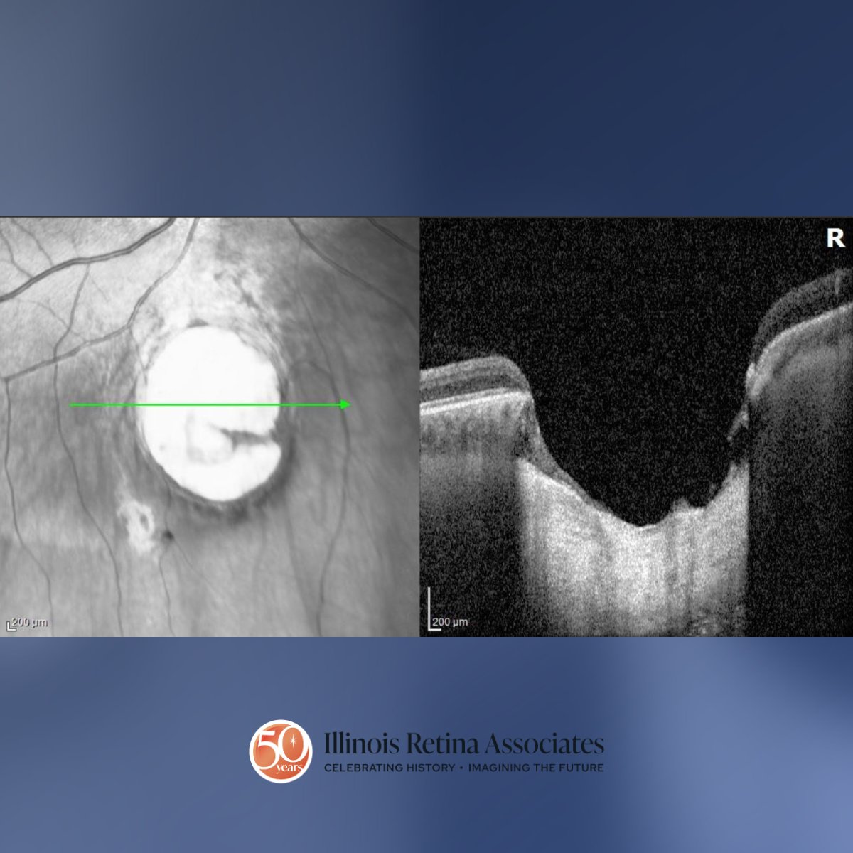
Visual acuity (VA) was 20/20 OU with correction. Intraocular pressures were normal in both eyes (OU). Anterior segment exam was unremarkable OU. Posterior segment exam in the right eye (OD) revealed a cup-to-disc ratio of 0.3, normal retinal vessels, unremarkable macula, and a well-defined 2-disc diameter circular area of atrophy with outpouching located 2-3-disc diameters inferior to the optic nerve (Fig 1). Posterior segment in the left eye (OS) was unremarkable. Ocular Coherence Tomography through this area OD revealed absence of retinal pigment epithelium and choroidal tissue with an outpouching into the sclera (Fig 2).

Differential Diagnosis:
- Chorioretinal coloboma
- Chorioretinal scar (i.e. ocular toxoplasmosis)
- Degenerative myopia
Discussion:
Chorioretinal Coloboma
Chorioretinal coloboma is a congenital abnormality caused by incomplete closure of the embryonic fissure during fetal development. It can be sporadic or inherited. The condition is characterized by absent retinal pigment epithelium and choroid in the affected area with a thin rudimentary neurosensory retinal layer often overlying the coloboma.1 It is commonly located inferior due to the inferonasal location of the embryonic fissure. If there is no involvement of the macula or optic nerve, patients are typically asymptomatic.1 It is important to monitor for retinal detachment as retinal breaks may form at the coloboma edge. Prophylactic laser photocoagulation can be considered around the coloboma.2 Occasionally, choroidal neovascular membranes may form near the coloboma and can be treated with anti-VEGF injections if active. Patients with systemic abnormalities in addition to coloboma or bilateral colobomas can be referred to a genetic specialist to evaluate for systemic disorders.3
If you are looking to schedule your first consultation, please contact us today by clicking HERE and find the location that is nearest you!
References:
- Fineman, M. S. (Ed.). (2024). Retina: Color Atlas and Synopsis of Clinical Ophthalmology (4th ed.). Wolters Kluwer. Lippincott Williams & Wilkins.
- Tripathy K, Chawla R, Sharma YR, Venkatesh P, et al. Prophylactic laser photocoagulation of fundal coloboma: does it really help? Acta Ophthalmol. 2016;94(8):e809-e810.
- Onwochei, B. C., Simon, J. W., Bateman, J. B., Couture, K. C. & Mir, E. Ocular colobomata. Surv. Ophthalmol. 45, 175–194 (2000).