Combined Branch Retinal Vein Occlusion And Branch Retinal Artery Occlusion – May, 2026
History:
A 69-year-old female with no known past medical history presented for sudden onset blurred vision in her left eye (OS) occurring 1 week prior. She denied floaters, flashes, total loss of vision, or headache.
Exam:
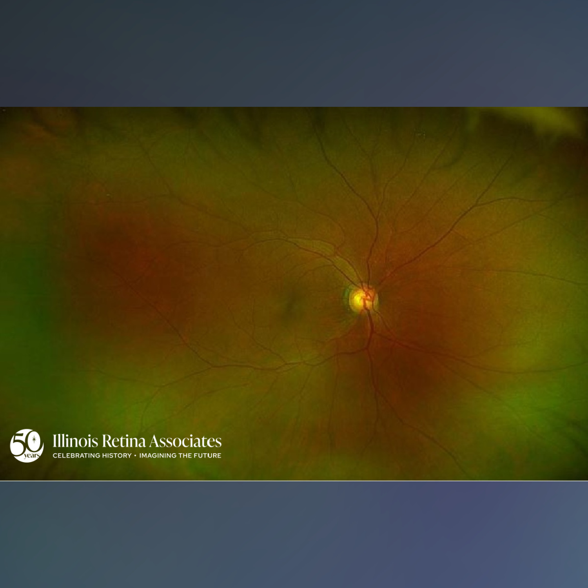
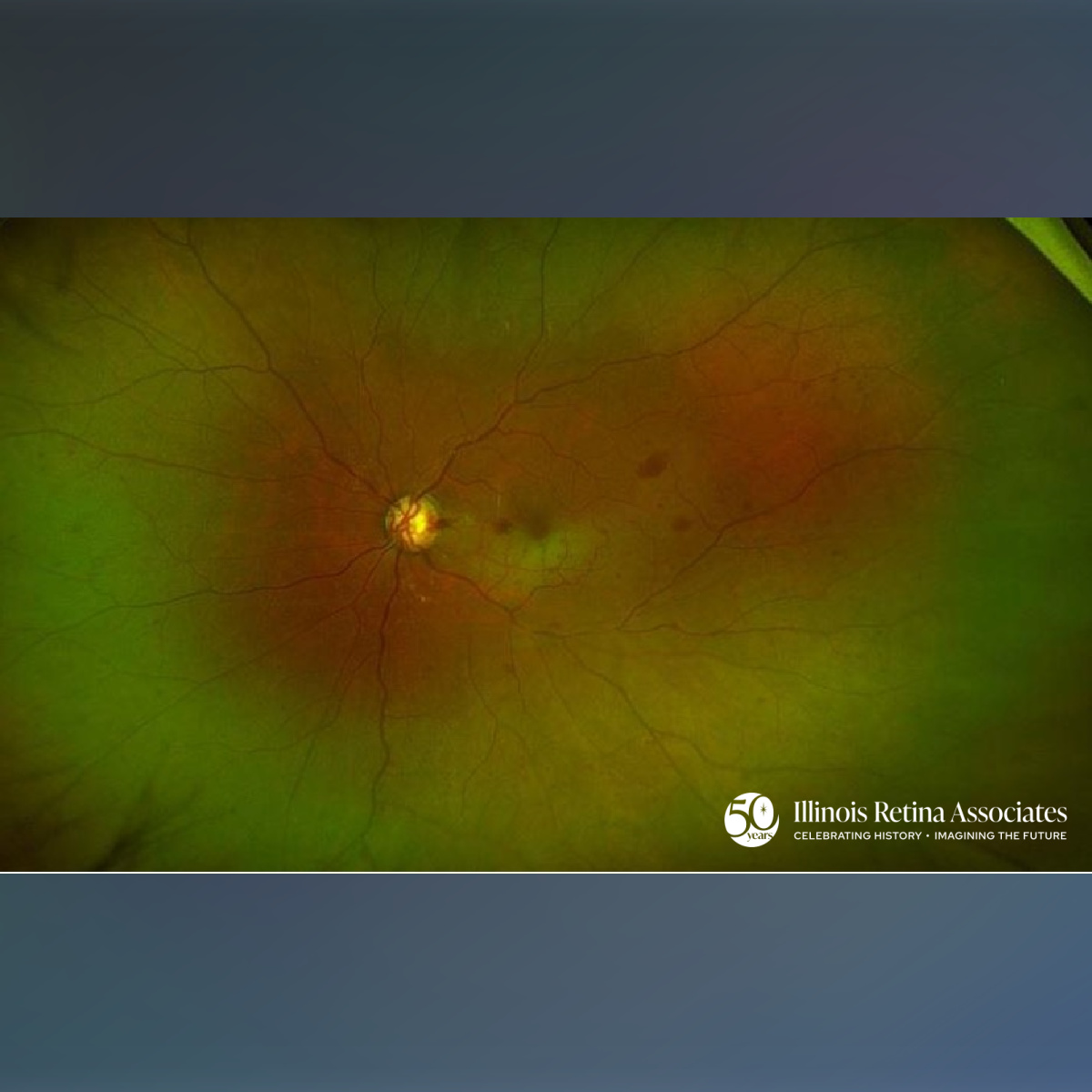
Visual acuity was 20/25 in the right eye (OD) and count fingers at 1 foot OS. Intraocular pressure was normal in both eyes (OU). Anterior segment exam OU was normal. Posterior segment exam OD demonstrated a cup-disc (c/d) ratioof 0.8, attenuated vessels, and AV nicking (Fig. 1). Posterior segment exam OS revealed a c/d ratio of 0.95, disc pallor, multiple disc hemorrhages, AV nicking, retinal whitening of the inferior macula, and scattered retinal hemorrhages (Fig. 2).
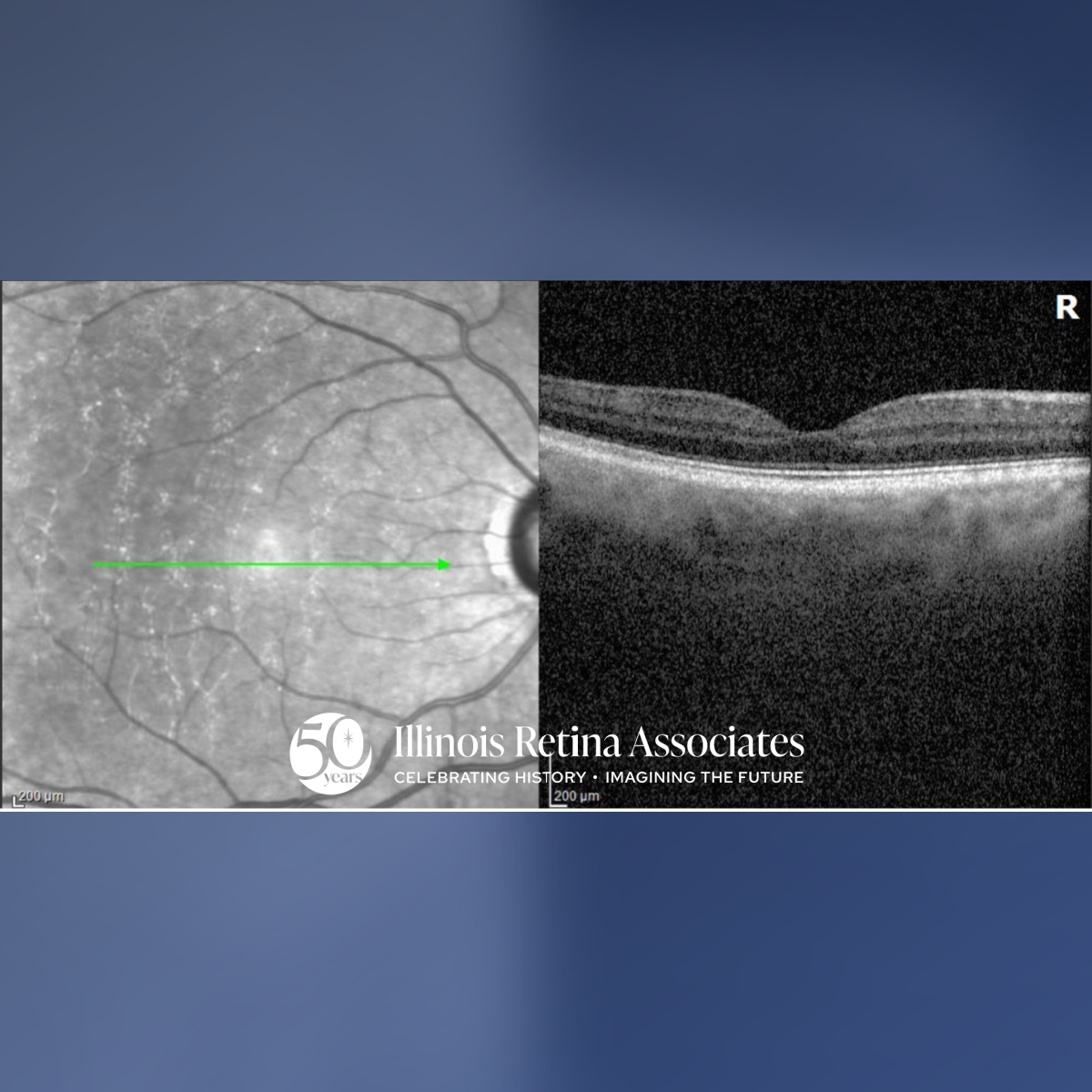
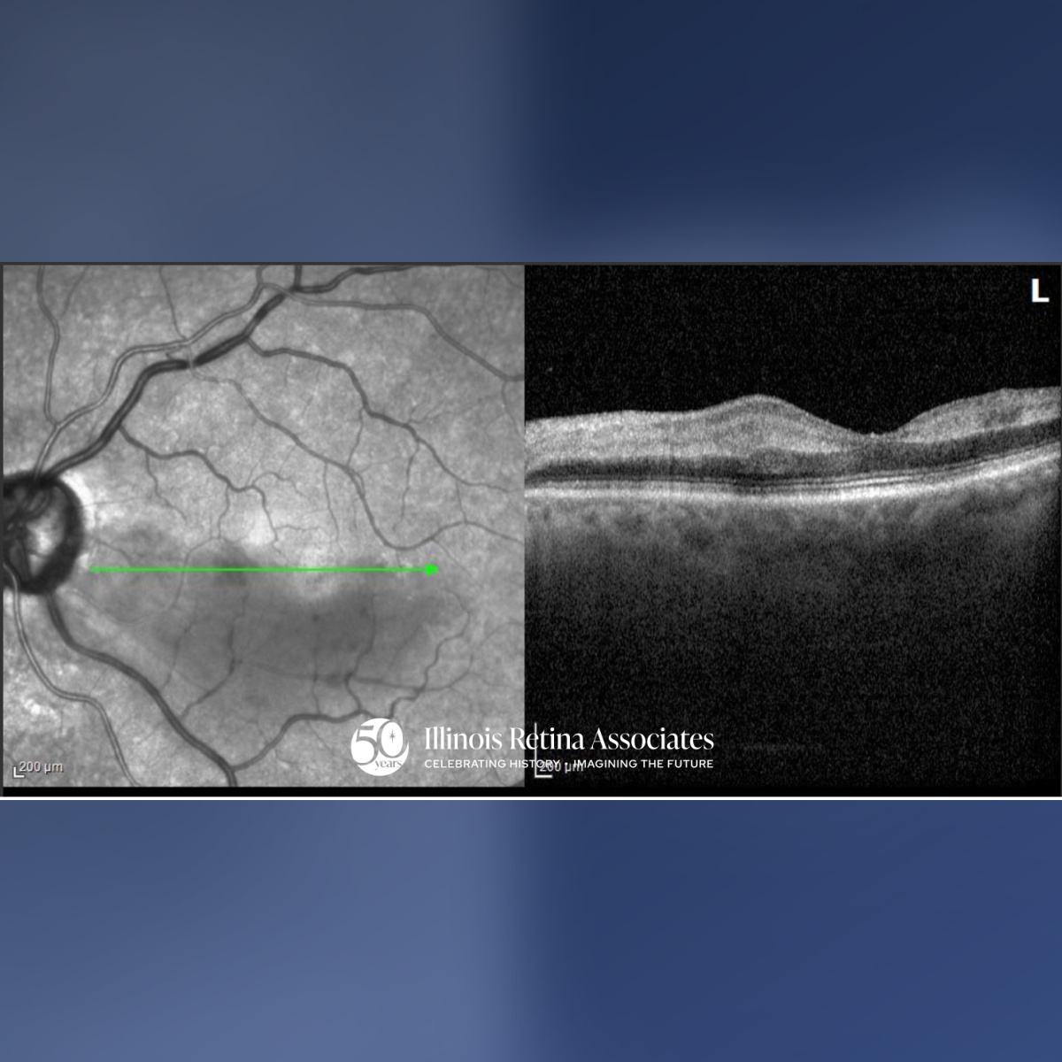
OCT OD was unremarkable (Fig. 3). OCT OS was significant for retinal edema and loss of inner retinal layer definition (Fig. 4). IVFA was performed which was normal OD but was notable for a delayed AV transit time, delayed arterial filling, and capillary nonperfusion over the inferior macula OS.
The patient was referred to the emergency department for urgent stroke evaluation at which point she was found to have a patent foramen ovale (PFO).
Differential Diagnosis of Central Retinal Artery Occlusion:
• Combined branch retinal vein occlusion and branch retinal artery occlusion (BRVO/BRAO)
• Isolated BRVO
• Embolic BRAO with secondary venous congestion
• Inflammatory Etiology/Retinal Vasculitis
Discussion:
Combined BRVO/BRAO:
Combined BRVO/BRAO is an uncommon retinal vascular event characterized by simultaneous or sequential involvement of both the arterial and venous circulation, often presenting with severe vision loss.1 These events can occur from an embolic etiology such as from paradoxical emboli in the setting of a PFO.2 Diagnosis involves exam with sectoral hemorrhages, retinal whitening, OCT demonstrating inner retinal hyperreflectivity consistent with ischemia, and IVFA revealing delayed arterial filling alongside venous congestion. Visual outcomes are variable and depend on the extent of ischemia.1,3
If you are looking to schedule your first consultation, please contact us today by clicking HERE and find the location that is nearest you!
References:
- Raval V, Nayak S, Saldanha M, Jalali S, Pappuru RR, Narayanan R, Das T. Combined retinal vascular occlusion: Demography, clinical features, visual outcome, systemic co-morbidities, and literature review. Indian J Ophthalmol. 2020 Oct;68(10):2136-2142. doi: 10.4103/ijo.IJO_2116_19. PMID: 32971625; PMCID: PMC7727993.
- Maiz AM, Murali S, Miller JML. Retinal artery occlusion in young patients without typical cardiovascular risk factors: etiologies, prognosis, and suggested work-up. Graefes Arch Clin Exp Ophthalmol. 2024 Nov;262(11):3577-3587. doi: 10.1007/s00417-024-06527-5. Epub 2024 Jun 7. PMID: 38847894.