Myopic Degeneration – January, 2026

History:
A 42-year-old patient with history of high myopia (-9D OU) presented with right eye (OD) central distortion for 1 day without pain or other visual changes. Patient denied history of trauma.
Exam:
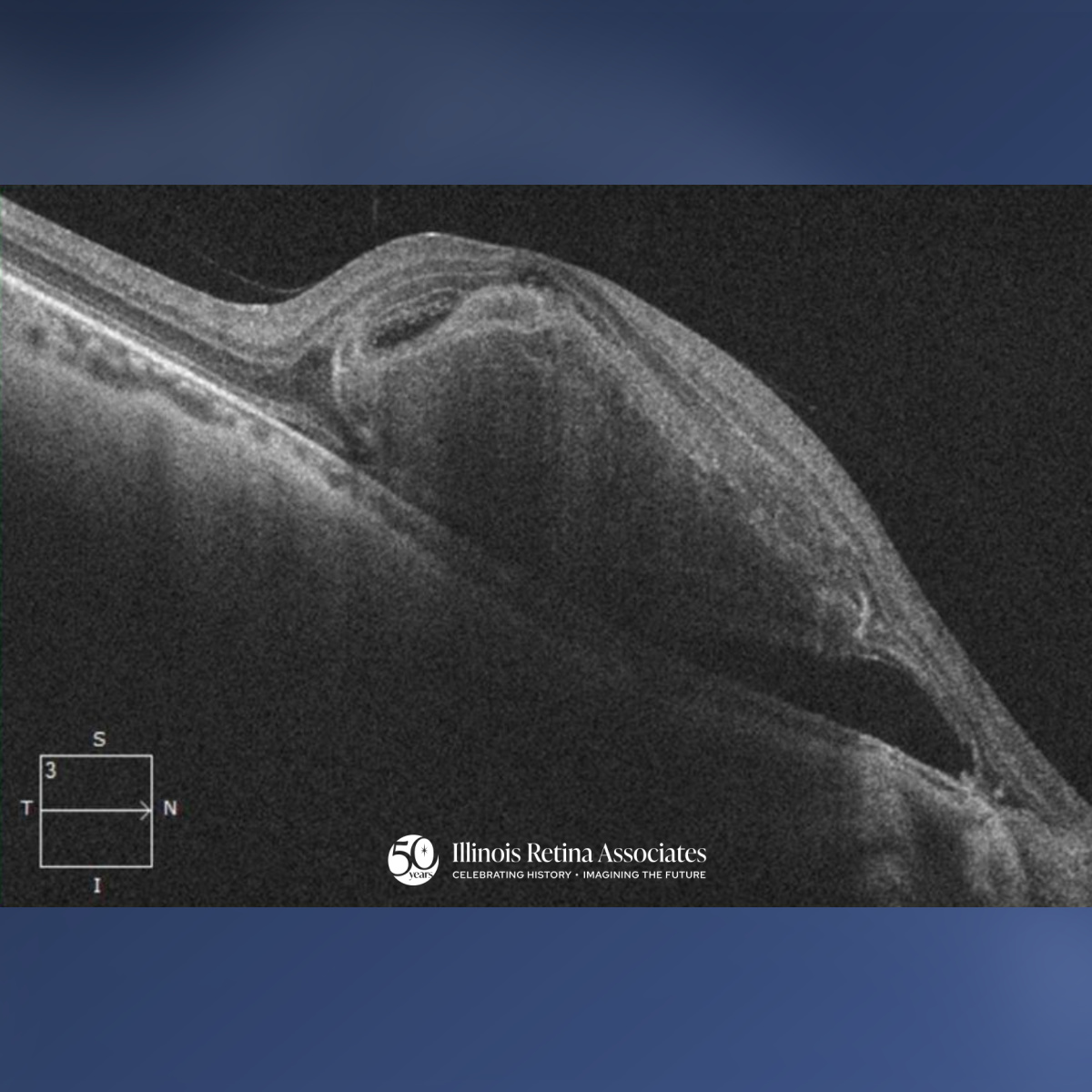
Visual Acuity was 20/150 OD and 20/60 pinhole 20/40 in the left eye (OS). Intraocular pressure was normal in both eyes (OU). Anterior segment was unremarkable OU. Posterior segment OD had a 0.2 CDR with peripapillary atrophy (PPA), vitreous syneresis, and a central macular subretinal hemorrhage with attached periphery. Posterior segment OS had a 0.4 CDR with PPA, posterior vitreous detachment, no macular hemorrhage or fluid, and attached periphery.

Differential Diagnosis:
- Presumed ocular histoplasmosis
- Myopic degeneration complicated by subretinal hemorrhage
- Wet age-related macular degeneration (AMD)
- Polypoidal choroidal vasculopathy
- Choroidal rupture
Discussion:
Mypopic Degeneration
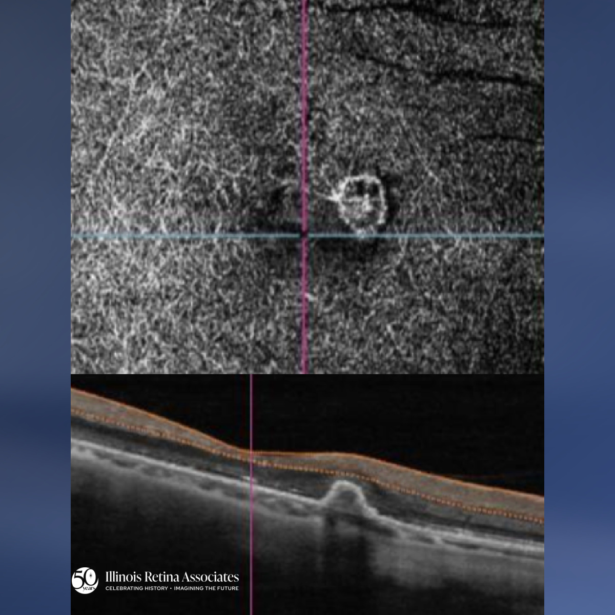
Myopic degeneration results from thinning of the retinal pigment epithelium and choroid leading to atrophy and possibly choroidal neovascularization (CNV).1 With this patient’s high myopia, she developed a large subretinal hemorrhage due to a lacquer crack (spontaneous linear break in Bruch membrane) with CNV. She received an intravitreal injection of Lucentis. Following 15 months of anti-VEGF injections, her subretinal fluid resolved however she still had a clear CNV membrane on OCT-angiography (Fig 3). CNV can develop in 5-10% of eyes with axial length >26.5mm and is often seen in association with lacquer cracks.1 Patients can also lose vision in degenerative myopia due to progressive macular atrophy.1 In contrast to AMD, CNV lesions in myopic degeneration are often more responsive to anti-VEGF and stable over time.
If you are looking to schedule your first consultation, please contact us today by clicking HERE and find the location that is nearest you!
References:
- Fineman, M. S. (Ed.). (2024). Retina: Color Atlas