Paracentral Acute Middle Maculopathy – February, 2026

History:
A 22-year-old male with a past medical history of hyperlipidemia presented for a sudden-onset “central grey spot” in his right eye (OD) occurring less than 24 hours prior to presentation. He denied new floaters or flashes, curtain over the vision, headache, or any neurological symptoms.
Exam:
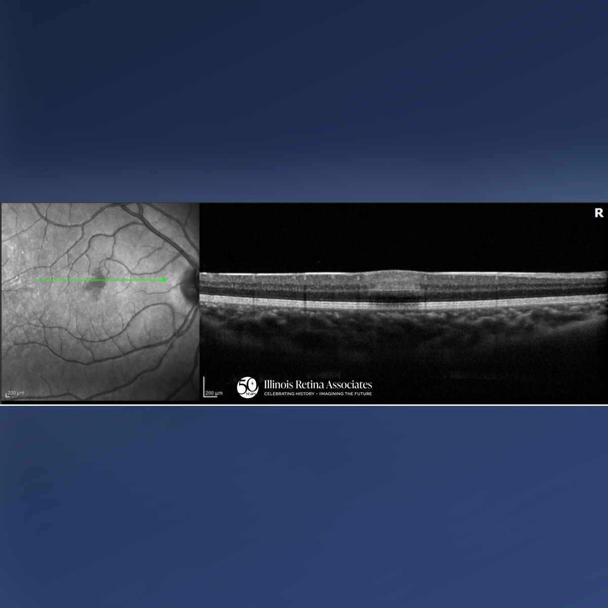
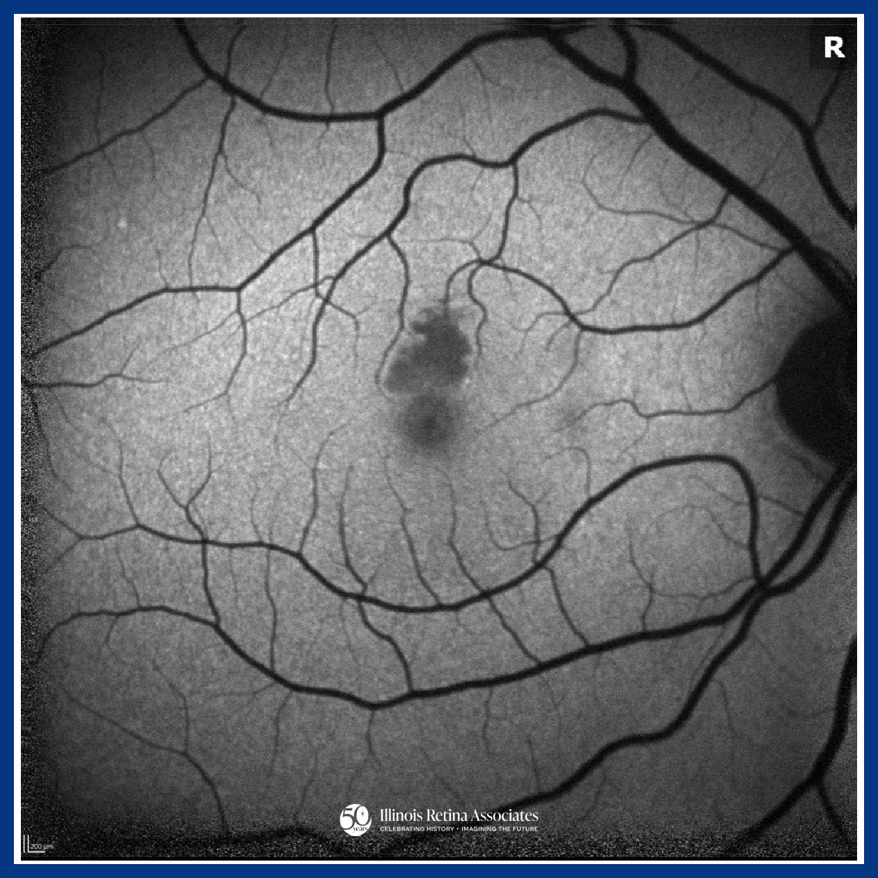
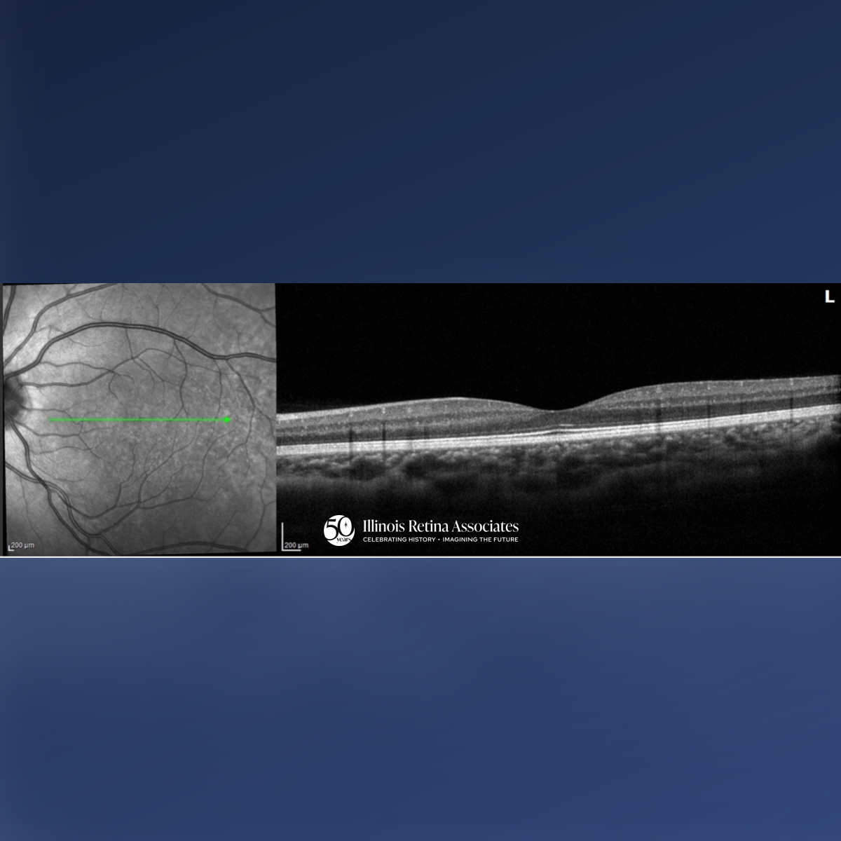
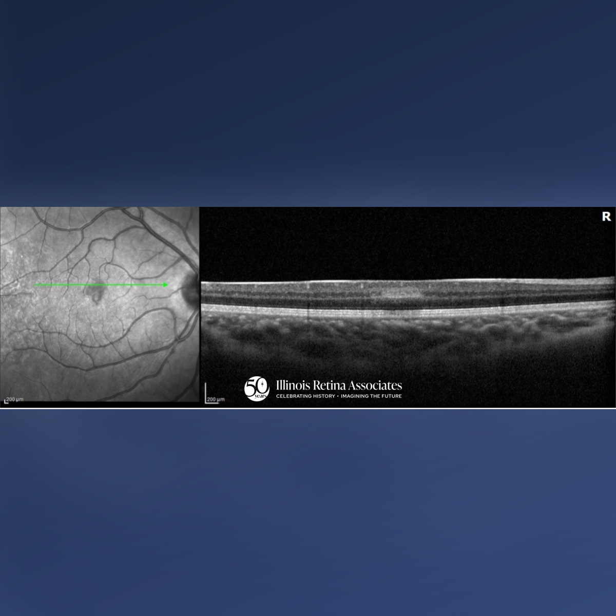
Visual acuity (VA) was 20/20 in both eyes (OU). Intraocular pressures and anterior segment exams were normal OU. Posterior segment exam OD demonstrated a cup to disc ratio (CDR) of 0.1 and a focal area of retinal whitening just superior to the fovea. Posterior segment in the left eye (OS) revealed a CDR of 0.1 and was otherwise unremarkable. Ocular Coherence Tomography (OCT) OD was significant for a band-like hyperreflectivity of the inner retinal layers (Fig 1) and FAF OD demonstrated mild hypo-autofluorescence overlying the area of retinal whitening (Fig 2). OCT OS was unremarkable (Fig 3). The patient was referred to the emergency department for a stroke evaluation which was negative. He returned approximately one week later where his VA remained stable at 20/20, and OCT showed slight improvement of the inner retinal hyperreflectivity (Fig 4). His central scotoma, however, remained unchanged.
Differential Diagnosis:
• Paracentral Acute Middle Maculopathy (PAMM)
• Acute Macular Neuroretinopathy (AMN)
• Retinal Artery Occlusion
Discussion:
Paracentral Acute Middle Maculopathy
PAMM is an ischemic macular disorder affecting the inner nuclear layer, often presenting with a sudden paracentral scotoma with minimal VA loss.1 PAMM can affect a wide-range of individuals from healthy patients to those with underlying systemic or retinal vascular disease, including arterial or venous occlusion, diabetes, or hypotension.2 Diagnosis is image-based: acute OCT imaging shows band-like hyperreflectivity of the inner nuclear layer, followed by focal thinning. FAF may be normal or show subtle hypo-autofluorescence. OCT-A can reveal capillary plexus flow deficits.3 While generally self-limited, paracentral scotomas may persist despite preserved visual acuity, reflecting permanent middle retinal ischemic injury.1
If you are looking to schedule your first consultation, please contact us today by clicking HERE and find the location that is nearest you!
References:
- Moura-Coelho N, Gaspar T, Ferreira JT, Dutra-Medeiros M, Cunha JP. Paracentral acute middle maculopathy-review of the literature. Graefes Arch Clin Exp Ophthalmol. 2020 Dec;258(12):2583-2596. doi: 10.1007/s00417-020-04826-1. Epub 2020 Jul 13. PMID: 32661700.
- Chen X, Rahimy E, Sergott RC, Nunes RP, Souza EC, Choudhry N, Cutler NE, Houston SK, Munk MR, Fawzi AA, Mehta S, Hubschman JP, Ho AC, Sarraf D. Spectrum of Retinal Vascular Diseases Associated With Paracentral Acute Middle Maculopathy. Am J Ophthalmol. 2015 Jul;160(1):26-34.e1. doi: 10.1016/j.ajo.2015.04.004. Epub 2015 Apr 4. PMID: 25849522.
- Rahimy E, Sarraf D. Paracentral acute middle maculopathy spectral-domain optical coherence tomography feature of deep capillary ischemia. Curr Opin Ophthalmol. 2014 May;25(3):207-12. doi: 10.1097/ICU.0000000000000045. PMID: 24614148.