Primary Vitreoretinal Lymphoma – September, 2025

History:
Patient is a 61-year-old female referred for changes in the retinal vasculature. Patient reported occasional floaters in both eyes (OU) and denied other symptoms.
Exam:
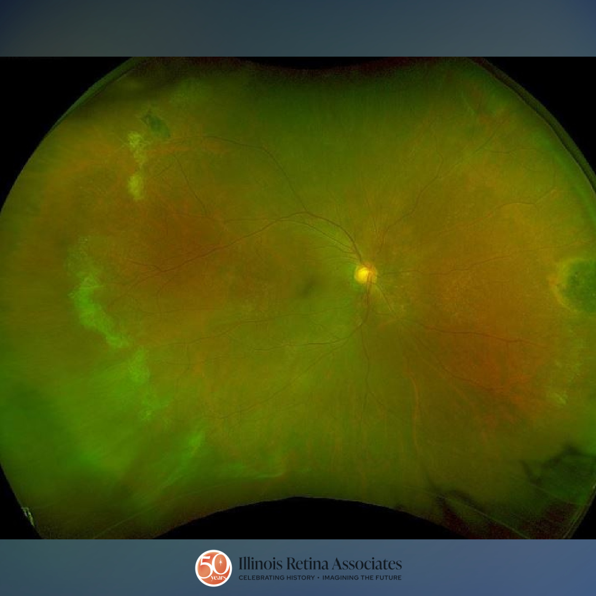
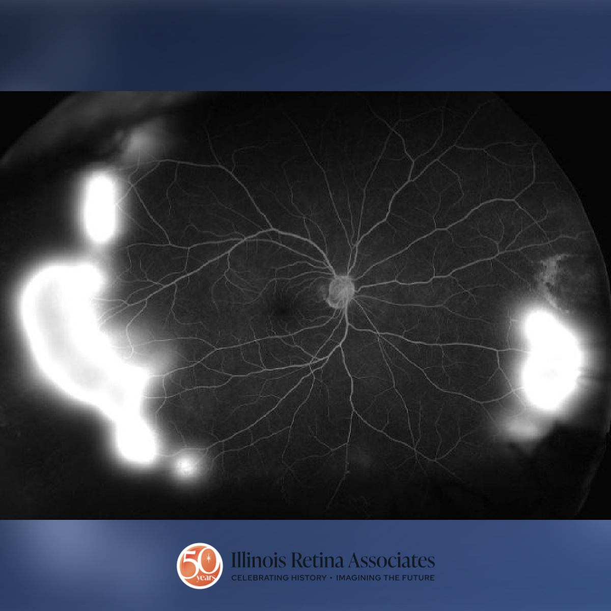
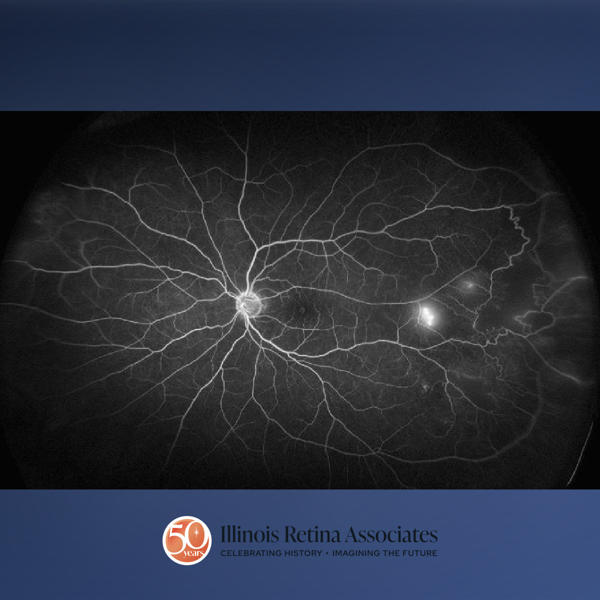
Visual Acuity (VA) was 20/30 pinhole 20/25 in the right eye (OD) and 20/40 pinhole 20/30 in the left eye (OS). Intraocular pressure (IOP) was normal OU and anterior segment exam was remarkable for mild cataracts OU. Posterior segment of the right eye revealed a 0.5 cup to disc ratio (CDR), normal macula, and numerous temporal and nasal peripheral sclerotic vascular seafan configurations and a nasal chorioretinal scar (Fig 1). The left eye showed 0.4 CDR and few peripheral sclerotic neovascular fronds. Fluorescein angiogram showed peripheral neovascularization worse OD (Fig 2) compared to OS (Fig 3) with peripheral nonperfusion.
Differential Diagnosis:
- Eales disease
- Proliferative diabetic retinopathy
- Radiation retinopathy
- Retinal vein occlusion
- Sarcoidosis
Discussion:
Primary Vitreoretinal Lymphoma
Sickle cell retinopathy is a disorder characterized by misshaped red blood cells obstructing retinal vasculature causing proliferative vascular changes. It is more common in those with sickle cell trait (SC) as opposed to the more systemically severe SS disease. Proliferative changes can result in neovascularization, vitreous hemorrhage, and retinal detachment. The disease is diagnosed clinically with fluorescein angiography aiding in assessing the severity. Treatment is typically not indicated for nonproliferative disease, though proliferative disease is often treated with panretinal photocoagulation to nonperfusion and surgery when indicated.
Upon questioning with our concern for sickle cell retinopathy, our patient reported she was sickle cell trait positive. She underwent panretinal photocoagulation areas of peripheral nonperfusion in both eyes.
If you are looking to schedule your first consultation, please contact us today by clicking HERE and find the location that is nearest you!
References:
- Fineman, M. S. (Ed.). (2024). Retina: Color Atlas and Synopsis of Clinical Ophthalmology (4th ed.). Wolters Kluwer. Lippincott Williams & Wilkins.