Leukemic Retinopathy – July, 2026

History:
A 49-year-old female with no known past medical history was referred for retinal hemorrhages in her right eye (OD). The patient endorsed seeing a “brown, smudge-like floater” starting 3 weeks prior.
Exam:
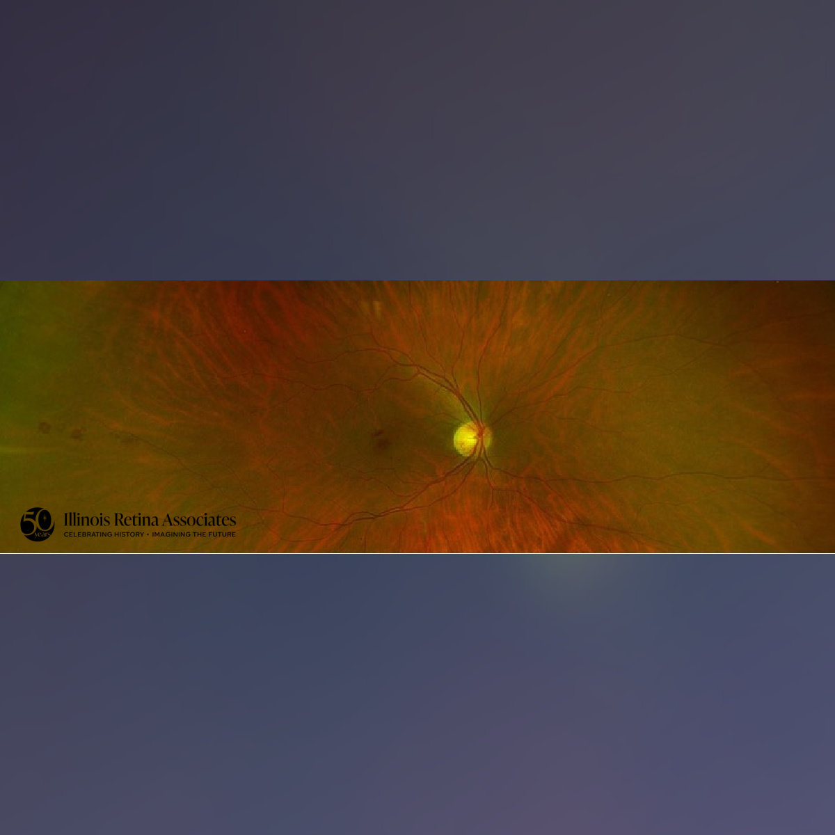
Visual acuity was 20/40 OD and 20/25 in the left eye (OS) and intraocular pressures were normal in both eyes (OU). There were no notable anterior segment findings OU. Posterior segment exam OD demonstrated intraretinal hemorrhage within the macula, scattered peripheral retinal hemorrhages, and two Roth spots temporally (Fig. 1). Posterior exam OS was unremarkable.
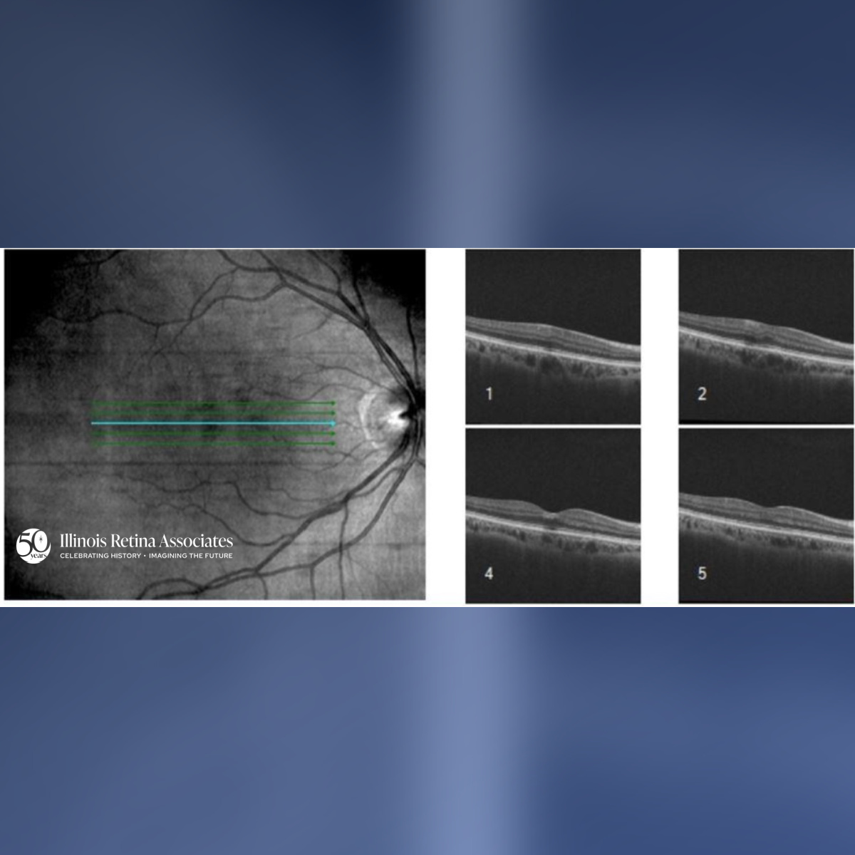
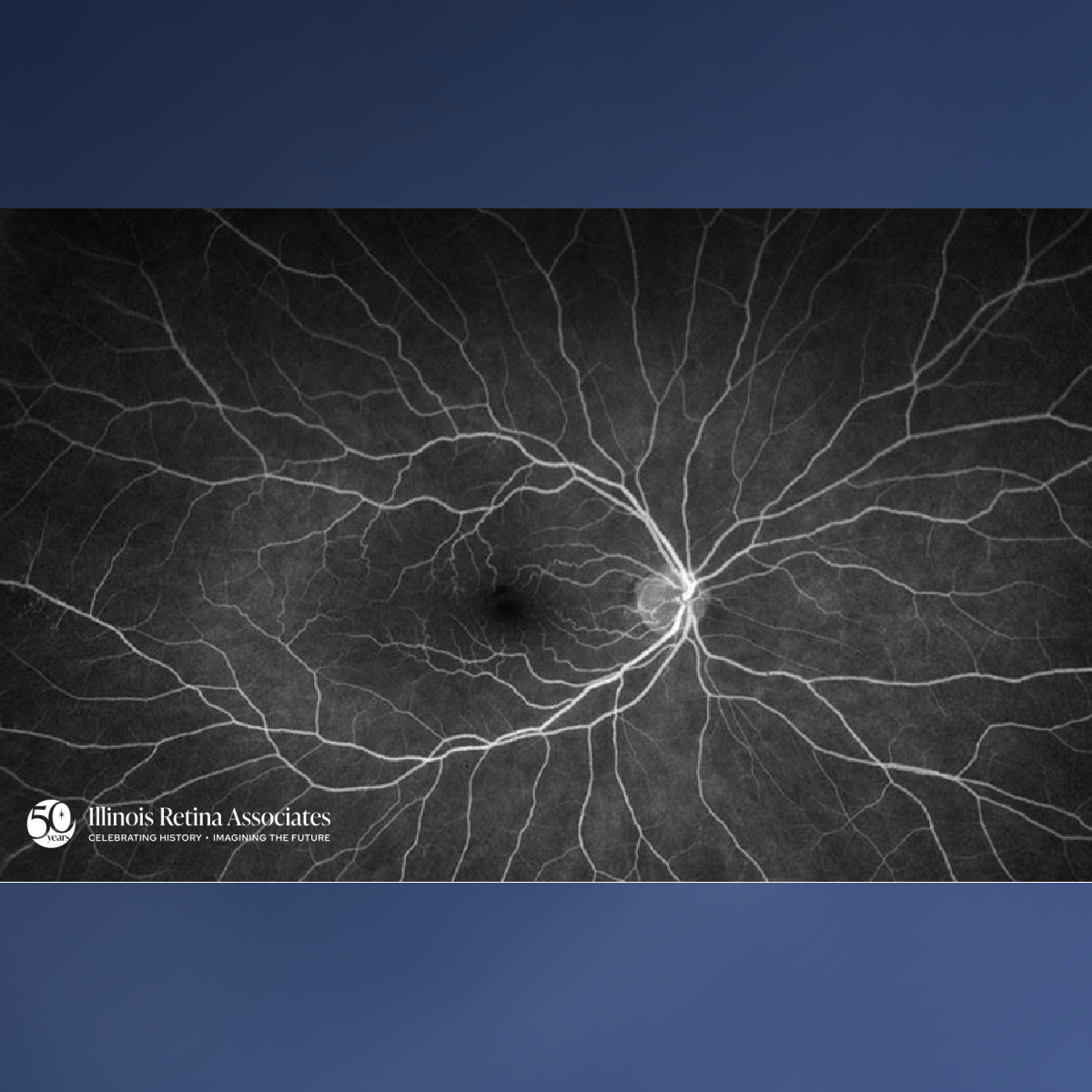
Optical Coherence Tomography (OCT) OD demonstrated mild inner retinal hyperreflectivity over the area of intraretinal hemorrhage (Fig. 2). OCT OS was unremarkable. IVFA was without neovascularization or ischemia OD (Fig 3) or OS.
Differential Diagnosis:
• Hypertensive Retinopathy
• Diabetic Retinopathy
• Anemia
• Hypercoagulable State
• Leukemia
• Carotid Stenosis
• Subacute Bacterial Endocarditis
Discussion:
Leukemic Retinopathy
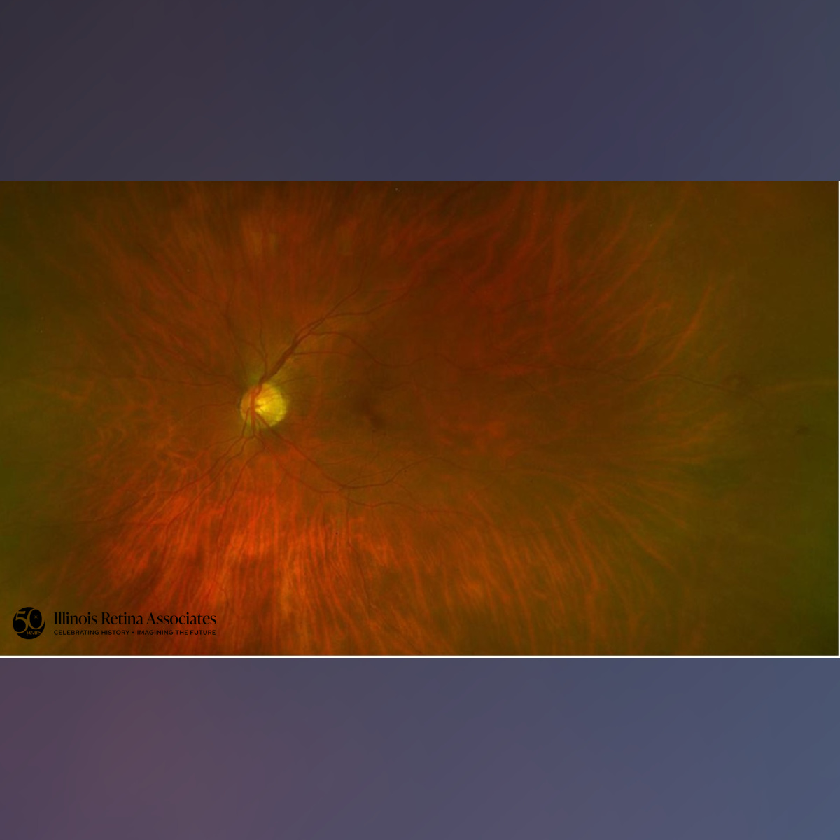
The patient was subsequently diagnosed with chronic lymphocytic leukemia (CLL). At her next visit she was found to have developed peripheral retinal hemorrhages OS (Fig 4). Leukemic retinopathy is a retinal vascular manifestation of systemic leukemia, characterized by indirect changes secondary to hematologic derangements.1 Such changes occur from a combination of anemia, thrombocytopenia, leukostasis, or hyperviscosity in the setting of bone marrow failure.1,2 Diagnosis involves findings of Roth spots (white-centered hemorrhages), multi-layered intraretinal hemorrhages, macular edema, inner retinal layer disruptions on OCT, or fluorescein angiography revealing areas of capillary nonperfusion alongside microaneurysms. Visual outcomes are variable and depend on the promptness of systemic chemotherapy and the successful regression of the underlying hematologic abnormalities.1,3
If you are looking to schedule your first consultation, please contact us today by clicking HERE and find the location that is nearest you!
References:
- Patel, N., Mohan, S., & Bhagat, N. (2025). Retinal Manifestations of Leukemia - Leukemic Retinopathy. EyeWiki. American Academy of Ophthalmology. https://eyewiki.org/Retinal_Manifestations_of_Leukemia_Leukemic_Retinopathy.
- Beketova, T., Mordechaev, E., Murillo, B., & Schlesinger, M. D. (2023). Leukemic Retinopathy: A Diagnostic Clue for Initial Detection and Prognosis of Leukemia. Cureus, 15(12). https://doi.org/10.7759/cureus.50587.
- Chandra, A., Chakraborty, U., Ganai, S., & Ray, A. K. (2020). Roth spots in acute myeloid leukaemia. BMJ Case Reports, 13(9), e238133. https://doi.org/10.1136/bcr-2020-238133.