Morning Glory Anomaly – October, 2025

History:
A 34-year-old female was referred for evaluation for longstanding limited vision in the left eye (OS).
Exam:
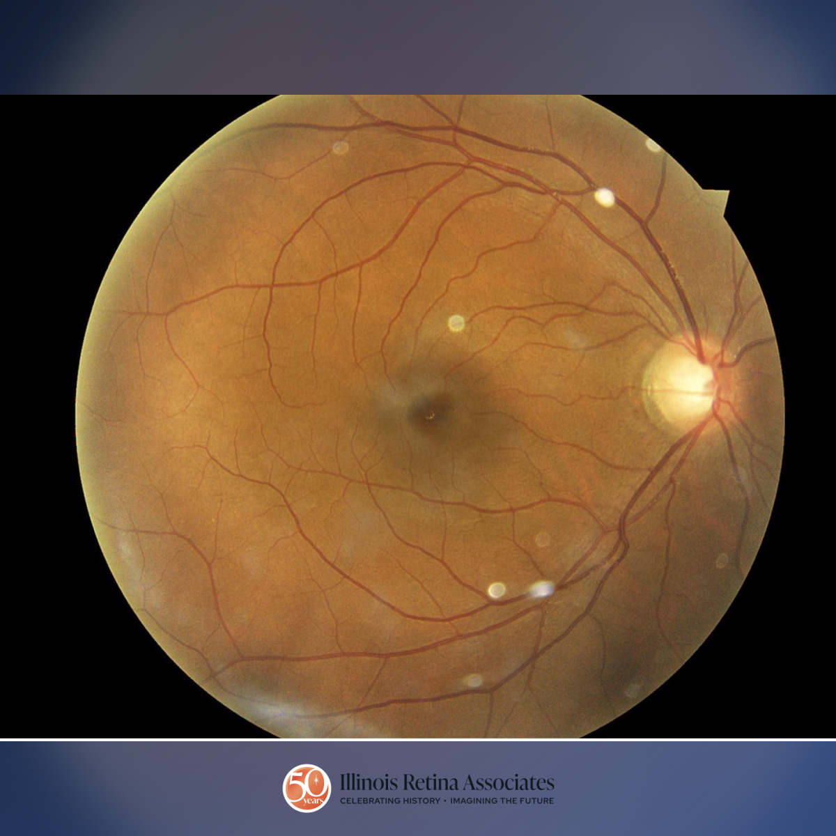
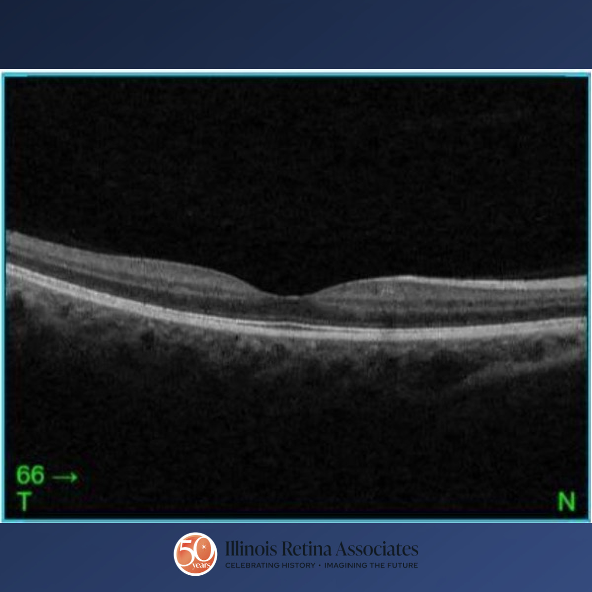
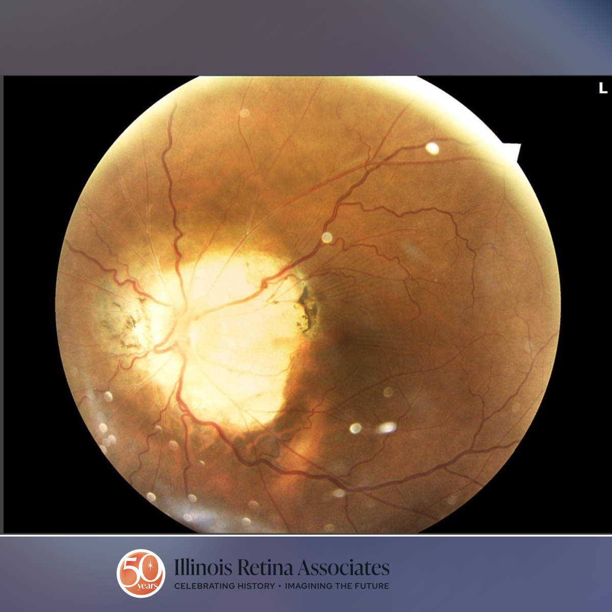
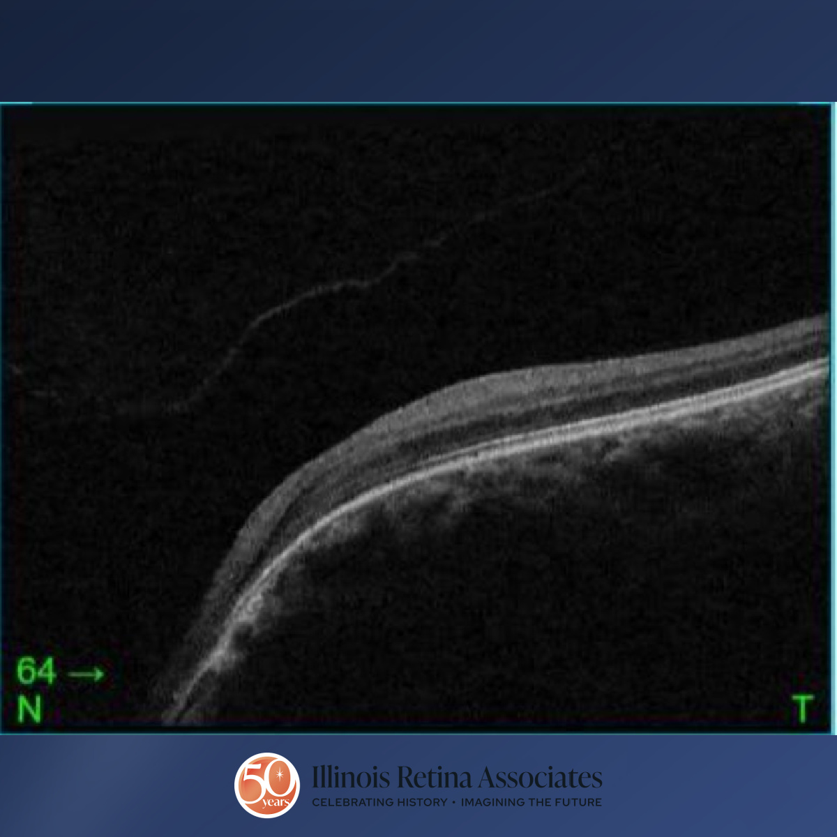
Visual acuity was 20/25 in the right eye (OD) and 20/40 OS. Intraocular pressures were within normal limits in both eyes (OU). Anterior segment exam was remarkable for persistent pupillary membranes bilaterally. Posterior segment exam OD demonstrated a cup to disc ratio of 0.5 and mild peripapillary atrophy (Fig 1) and OCT demonstrated vitreomacular adhesion (Fig 2). Posterior segment exam OS revealed a funnel-shaped optic nerve with a c/d ratio of 0.5, peripapillary atrophy and pigmentation, and straightened retinal blood vessels overlying the optic disc margin (Fig 3). OCT OS was significant for a nasal staphyloma (Fig 4). A previous MRI/MRA of the brain was normal.
Differential Diagnosis:
• Morning Glory Anomaly
• Optic Nerve Coloboma
• Peripapillary Staphyloma
Discussion:
Morning Glory Anomaly
Morning glory anomaly is a rare condition resulting from a congenital malformation of the optic nerve. It is typically more common in females and is theorized to occur from a deficit in embryonic fissure closure or primary mesenchymal differentiation during fetal development.1 When associated with systemic findings, such as intracranial structural or cerebrovascular abnormalities, it is referred to as morning glory syndrome.
Exam findings typically include a large, funnel-shaped, and excavated optic nerve head which resembles a morning glory flower.2 Additionally, patients may have peripapillary atrophy as well as numerous straight retinal vessels overlying the optic disc margin. Some patients may develop serous maculopathy.3
Visual acuity can range from normal to significantly limited and usually corresponds to the extent of the optic nerve malformation. Patients typically have visual field defects including enlarged blind spots.4
While there is no treatment for morning glory anomaly itself, patients should have brain imaging to assess for structural or vascular intracranial malformations that may need to be addressed surgically.2
If you are looking to schedule your first consultation, please contact us today by clicking HERE and find the location that is nearest you!
References:
- Cennamo, G et al. 2009. Evaluation of Morning Glory Syndrome with Spectral Optical Coherence Tomography and Echography. Ophthalmology. 117:6, p1269–1273.
- Lee, BJ and Traboulsi, EI. 2008. Update on the Morning Glory Disc Anomaly. Ophthalmic Genetics 29:2, p47-52.
- Brodsky, M.C. 2010. Congenital Optic Disc Anomalies in Pediatric Neuro-ophthalmology. 2nd ed. New York: Springer
- Brodsky, MC. 1994. Congenital Disc Anomalies. Survey of Ophthalmology 39:2, p89-112.