Coats Disease – November, 2026
History:
A 31-year-old male with no significant past medical history was referred for a suspected macula-on retinal detachment in the right eye (OD).
Exam:
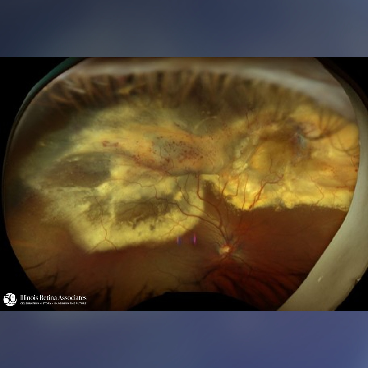
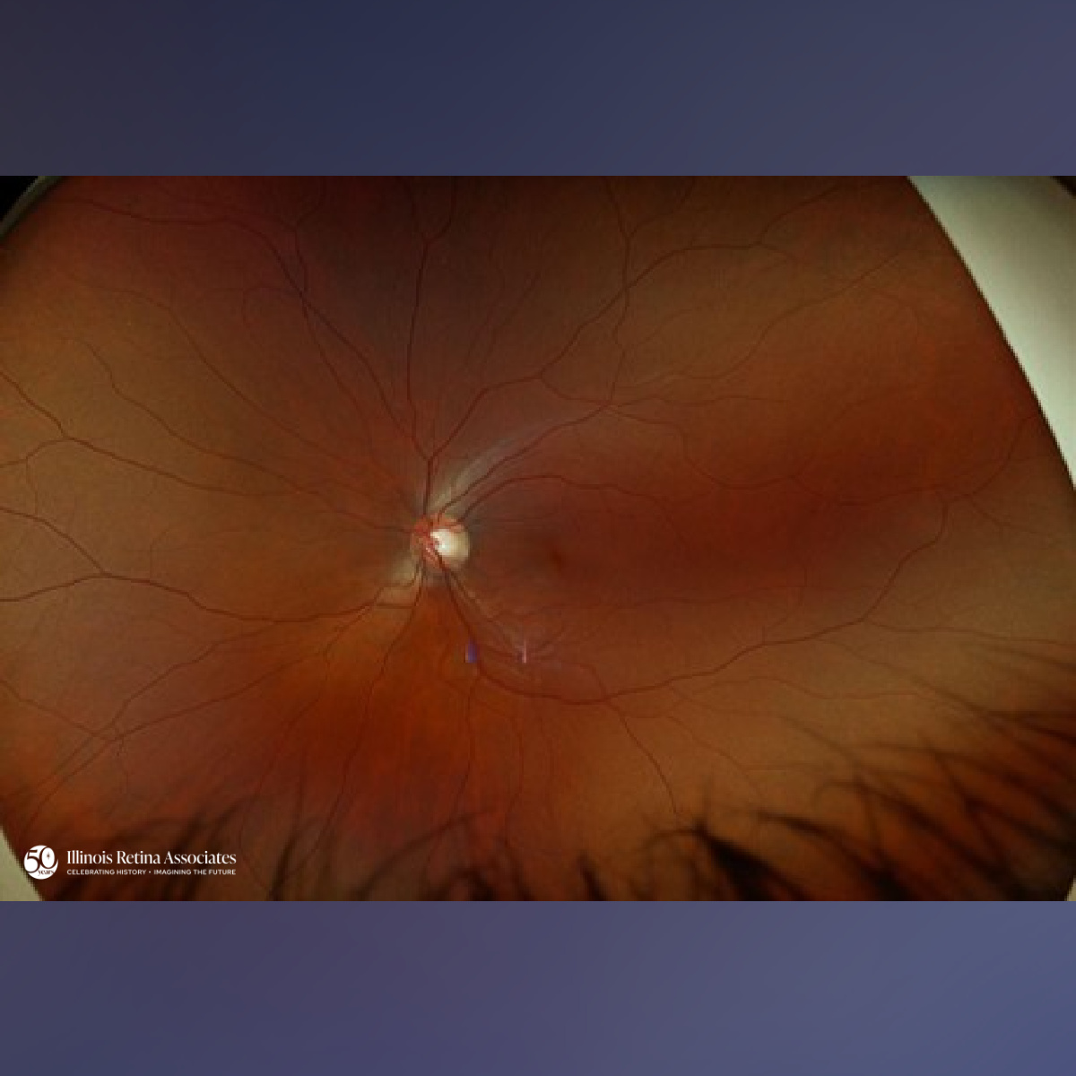
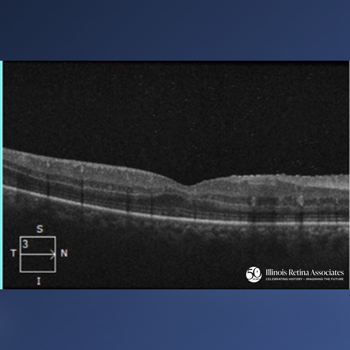
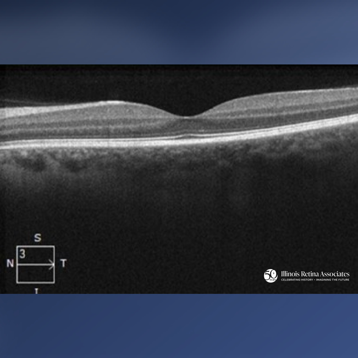
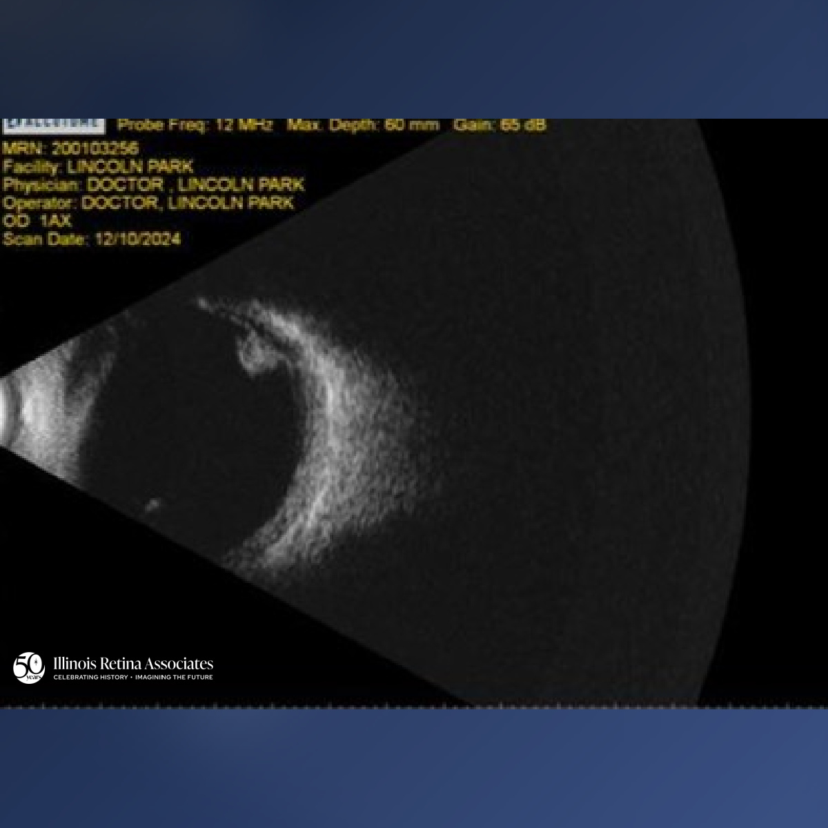
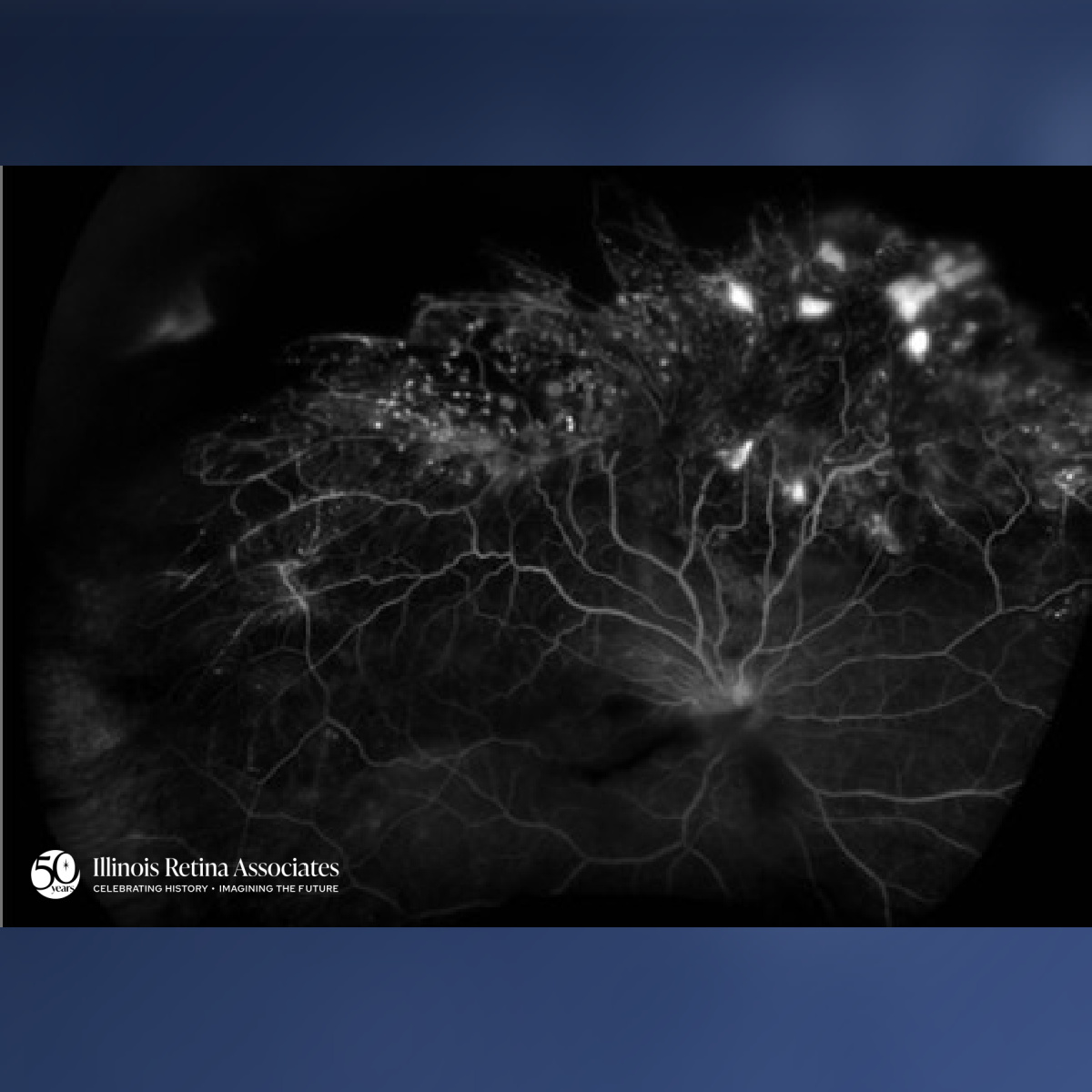
Visual acuity was 20/30 OD and 20/20 in the left eye (OS). Anterior segment was unremarkable in both eyes (OU). Dilated fundus exam OD demonstrated extensive peripheral retinal telangiectasia, aneurysmal vessels, intraretinal hemorrhages, and confluent yellow lipid exudation involving the superior and temporal retina, with associated retinal elevation concerning for an exudative retinal detachment (Fig 1). Fundus examination OS was unremarkable (Fig 2). Optical Coherence Tomography (OCT) OD showed mild inner retinal irregularity without macular fluid (Fig 3), while OCT OS was normal (Fig 4). B-scan ultrasonography confirmed a shallow exudative retinal detachment with retinal thickening (Fig 5). Fluorescein angiography revealed extensive telangiectasia, light-bulb aneurysms, vascular leakage, and peripheral capillary nonperfusion OD (Fig 6).
Differential Diagnosis:
• Coats disease
• Retinal vasoproliferative tumor
• Familial exudative vitreoretinopathy
• Retinal capillary hemangioblastoma
• Peripheral exudative hemorrhagic chorioretinopathy
Discussion:
Coats Disease
The findings were consistent with Coats disease, an idiopathic, predominantly unilateral retinal vascular disorder characterized by telangiectasia and progressive lipid exudation. Although typically diagnosed in young males during childhood, adult-onset disease is recognized and often follows a more indolent course.
Abnormal retinal vascular permeability leads to leakage of plasma-derived lipoproteins, resulting in intraretinal and subretinal exudation and, in advanced cases, exudative retinal detachment. Treatment focuses on ablating abnormal vessels with laser photocoagulation or cryotherapy. Anti-VEGF therapy may be used as an adjunct in selected cases. Visual prognosis depends primarily on macular involvement and chronicity of exudation. In this patient, preserved foveal anatomy and good presenting visual acuity were favorable prognostic features.
If you are looking to schedule your first consultation, please contact us today by clicking HERE and find the location that is nearest you!
References:
-
Sen M, Shields C, Honavar S, Shields J. Coats disease: An overview of classification, management and outcomes. Indian J Ophthalmol. 2019;67(6):763. doi:10.4103/ijo.IJO_841_19
-
Ghorbanian S, Jaulim A, Chatziralli IP. Diagnosis and Treatment of Coats’ Disease: A Review of the Literature. Ophthalmologica. 2012;227(4):175-182. doi:10.1159/000336906