Myopic Foveoschisis – September, 2026

History:
A 65-year-old male with HTN, DVT, and myopia was referred for 6 months of worsening right-eye (OD) vision starting 6 months prior to presentation.
Exam:
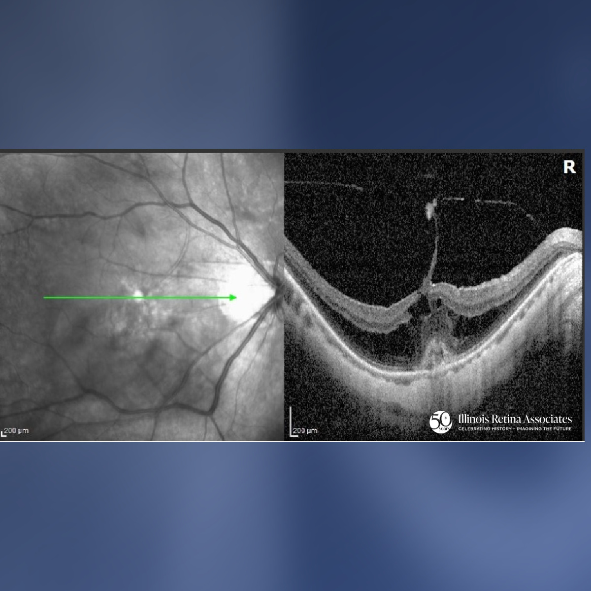
Visual Acuity was 20/70 OD, 20/30 in the left eye (OS); Intraocular pressures normal in both eyes (OU). Anterior segment: mild cataracts OU. Posterior segment exam OD revealed posterior staphyloma, central macular subretinal hemorrhage, mild ERM, foveoschisis, and treated peripheral lattice degeneration. Posterior segment exam OS showed posterior staphyloma, mild ERM, and a treated peripheral retinal tear.
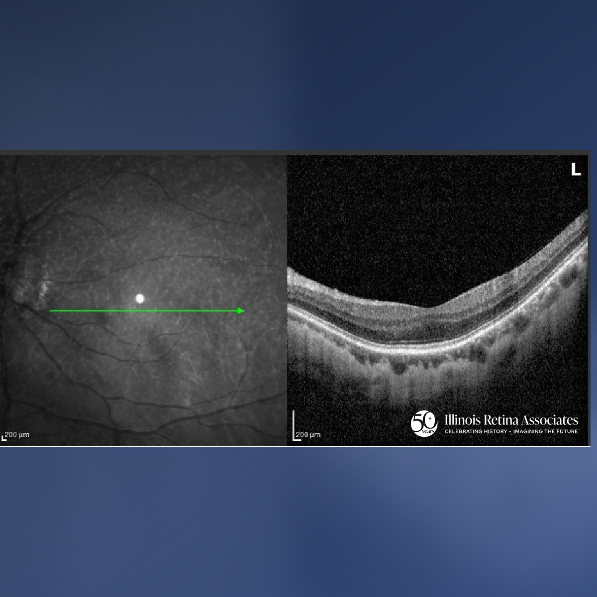
Optical Coherence Tomography (OCT) OD was significant for foveoschisis, mild ERM, and subretinal hyperreflective changes consistent with subretinal hemorrhage over an underlying myopic CNVM (Fig 1). OCT OS showed mild ERM and no hemorrhage or fluid (Fig 2).
Differential Diagnosis:
• Myopic foveoschisis (myopic traction maculopathy)
• Tractional foveoschisis from ERM or VMT
• Myopic macular hemorrhage from lacquer cracks
• Subretinal hemorrhage from neovascular AMD vs. myopic CNVM
Discussion:
Myopic Foveoschisis
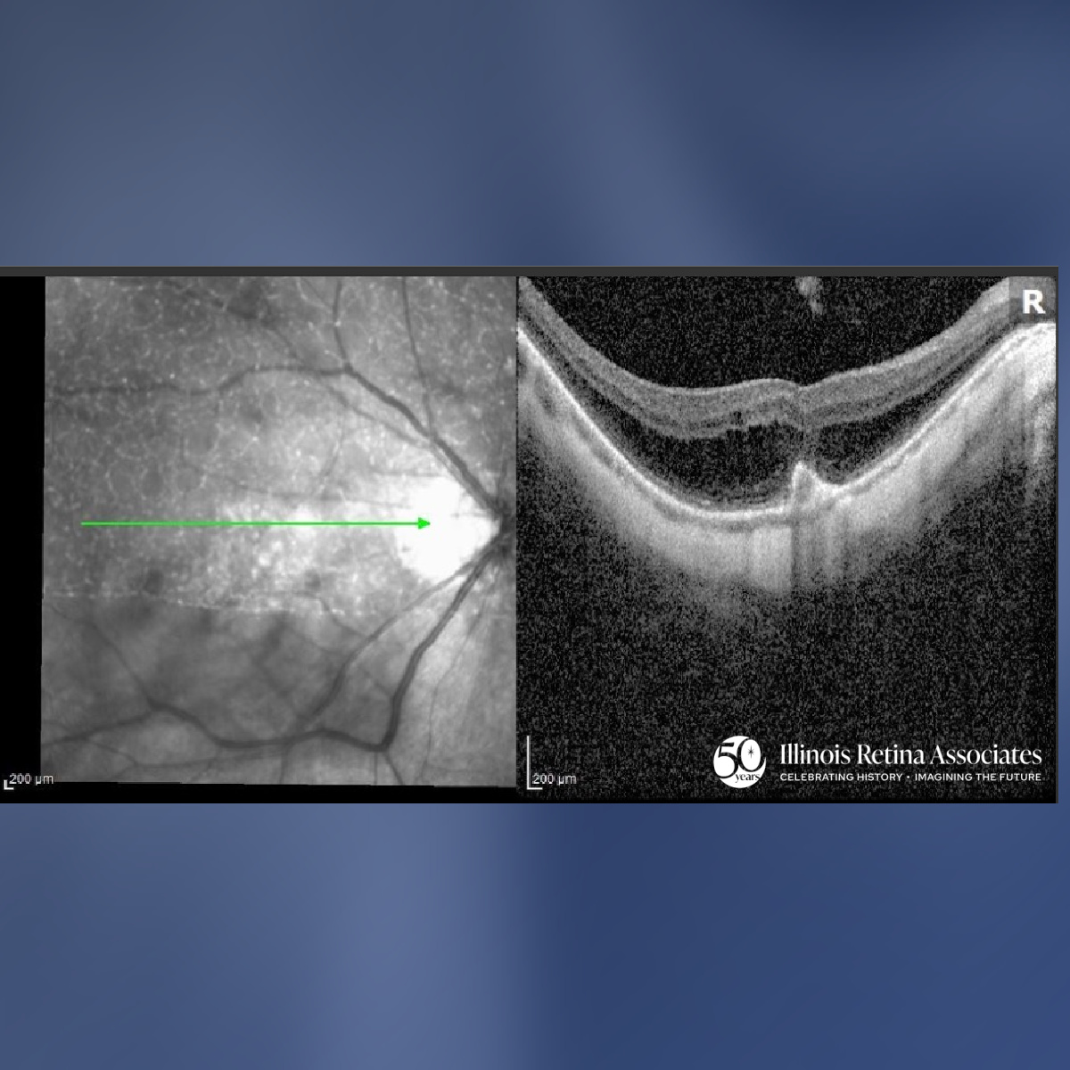
Based on the OCT appearance OD, the patient was diagnosed with myopic foveoschisis and subretinal hemorrhage from a myopic CNVM. After discussion, he elected to proceed with anti-VEGF injections. At 4 weeks, vision improved slightly to 20/70 with improving hemorrhage on exam and OCT (Fig 3).
Myopic foveoschisis is a tractional maculopathy of highly myopic eyes, marked by splitting of the neurosensory retina into inner and outer layers. It results from posterior staphyloma, epiretinal tangential traction, internal limiting membrane rigidity, and retinal arteriolar traction amid progressive axial elongation.1 It may also arise in the setting of myopic CNV with subretinal hemorrhage.2 Diagnosis relies on OCT showing outer retinoschisis with columnar bridging elements, ILM detachment, and secondary foveal detachment or macular hole. Visual outcomes vary with photoreceptor and Henle fiber integrity and timely vitrectomy with ILM peeling.3
If you are looking to schedule your first consultation, please contact us today by clicking HERE and find the location that is nearest you!
References:
- Wu PC, Chen YJ, Chen YH, Chen CH, Shin SJ, Tsai CL, Kuo HK. Factors associated with foveoschisis and foveal detachment without macular hole in high myopia. Eye (Lond). 2009 Feb;23(2):356-61. doi: 10.1038/sj.eye.6703038. Epub 2007 Dec 7. PMID: 18064059.
- Shimada N, Ohno-Matsui K, Yoshida T, Futagami S, Tokoro T, Mochizuki M. Development of macular hole and macular retinoschisis in eyes with myopic choroidal neovascularization. Am J Ophthalmol. 2008 Jan;145(1):155-161. doi: 10.1016/j.ajo.2007.08.029. Epub 2007 Nov 7. PMID: 17988641.
- Sborgia G, Boscia F, Niro A, Giancipoli E, D'Amico Ricci G, Sborgia A, Sborgia L, Recchimurzo N, Romano MR, Addabbo G, Alessio G. Morphologic and functional outcomes of different optical coherence tomography patterns of myopic foveoschisis after vitrectomy and inner limiting membrane peeling. Eye (Lond). 2019 Nov;33(11):1768-1775. doi: 10.1038/s41433-019-0490-3. Epub 2019 Jun 17. PMID: 31209260; PMCID: PMC7002739.